If you have been told that you have a degenerative disc, bulging disc or herniated disc, it is easy to imagine the disc as a worn-out cushion that can only continue to deteriorate.
The biology is considerably more interesting than that.
Intervertebral discs are living tissues. They contain cells, consume nutrients, produce and maintain an extracellular matrix, respond to mechanical loading and undergo biological changes throughout life.
That doesn’t mean every damaged disc can simply “grow back” or return to its original condition. Disc degeneration can involve structural changes that may be permanent.
But research increasingly suggests that the intervertebral disc is not simply an inert structure destined to wear out.
To understand what recovery might be possible, we first have to understand how a spinal disc stays alive.
Between most of the vertebrae in the spine sits an intervertebral disc.
Each disc has three important components:
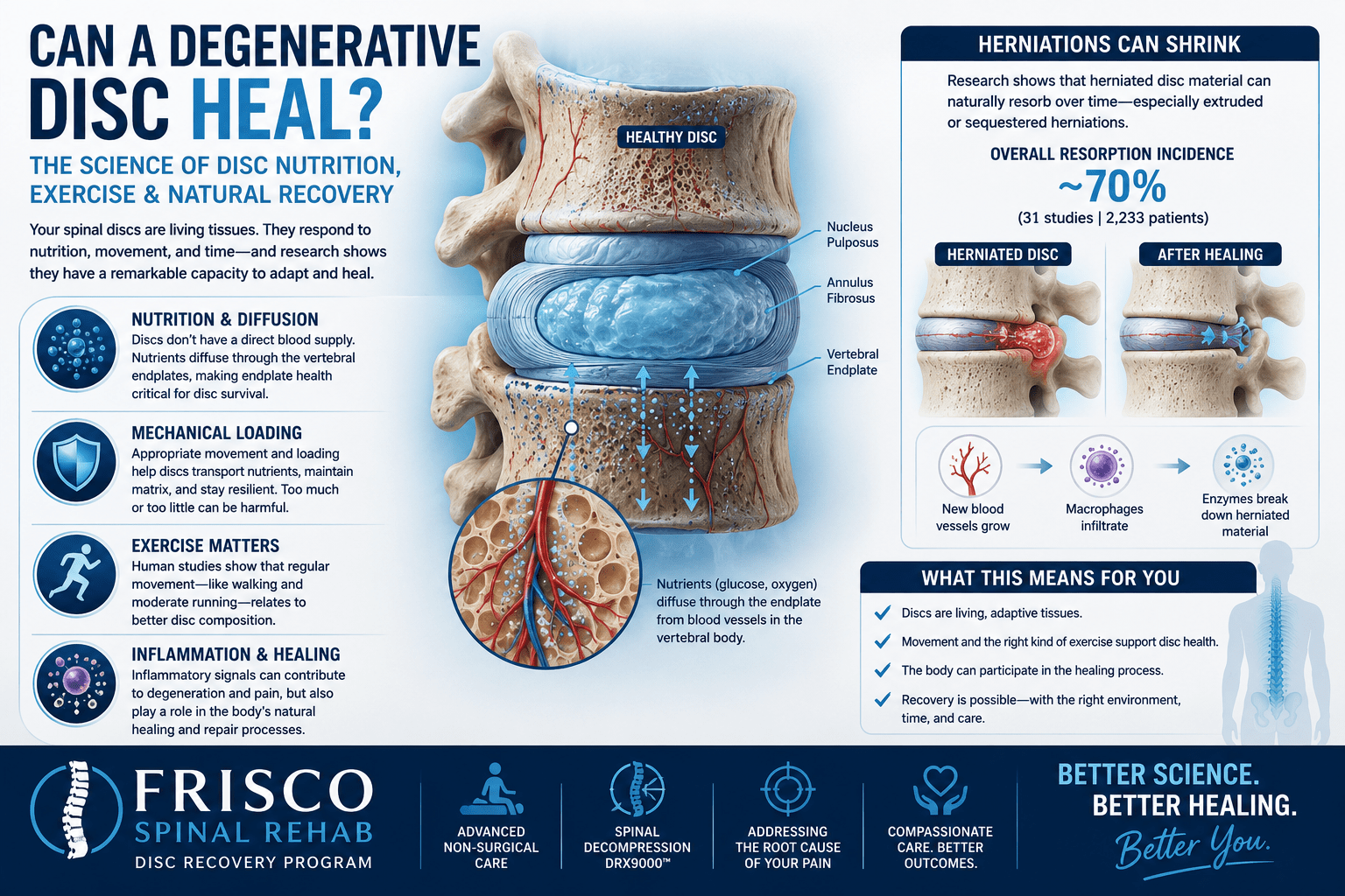
The nucleus pulposus is the softer, highly hydrated central portion of the disc.
The annulus fibrosus is the strong, layered outer ring that surrounds and contains the nucleus.
The vertebral endplates are thin layers separating the disc from the vertebral bodies above and below it.
Together, these structures allow the spine to tolerate compression, bending and movement while distributing forces between the vertebrae.
One of the most fascinating things about the disc, however, is something it largely doesn’t have: a direct blood supply.
Most tissues receive oxygen and nutrients directly through blood vessels.
The adult intervertebral disc is different.
The inner portion of a healthy adult disc is largely avascular, meaning that blood vessels do not directly penetrate most of the tissue. Only the outermost annulus has a limited vascular supply.
So how do the cells deep inside a disc survive?
Primarily through transport of nutrients from blood vessels adjacent to the vertebral endplates and, to a lesser extent, the outer annulus.
Small molecules such as glucose and oxygen move primarily by diffusion.
This is important because disc cells still require nutrients to remain viable and maintain the extracellular matrix even though those nutrients have to travel considerable distances to reach them.
Researchers have described steep gradients across the disc: oxygen and glucose tend to become lower toward the center while metabolic byproducts such as lactate increase.
The center of the disc therefore exists in an unusually low-oxygen, relatively acidic biological environment.
Disc cells are adapted to this environment—but there are limits.
If nutrient delivery becomes inadequate, the cells may have difficulty maintaining healthy disc tissue.
When people look at a lumbar MRI, they naturally focus on the discs.
But the vertebral endplates immediately above and below the disc are critically important to disc biology.
Blood vessels in the vertebral bodies bring nutrients close to these endplates. Nutrients can then move across the endplate and into the disc.
The endplates therefore function somewhat like an exchange interface between the body’s circulation and the relatively avascular disc.
Changes associated with aging, sclerosis, calcification or injury can potentially reduce endplate permeability.
If that happens, nutrient transport into the disc and removal of metabolic waste may become less efficient.
This has led researchers to investigate impaired disc nutrition as one potential contributor to the degenerative process.
Disc degeneration is much more complicated than simple “wear and tear.”
A healthy nucleus pulposus contains molecules called proteoglycans that attract and retain water.
That water is important.
It helps the disc resist compression and distribute mechanical forces.
As degeneration progresses, the biochemical composition of the disc can change. Proteoglycan content may decline, water content can decrease and the extracellular matrix can become increasingly disorganized.
The disc may consequently lose height and some of its ability to distribute loads.
Meanwhile, changes can occur in the annulus fibrosus, including fissures and disruption of its organized collagen structure.
The result can be the familiar MRI descriptions patients frequently see:
But the MRI is showing structure, not the complete biological story.
Degenerated and herniated discs can contain increased levels of inflammatory signaling molecules, including cytokines such as tumor necrosis factor-alpha and several interleukins.
These inflammatory pathways may contribute both to degradation of the disc’s extracellular matrix and to pain.
This helps explain an important clinical observation:
The size of a disc abnormality on MRI doesn’t always correspond neatly with how much pain a patient experiences.
Mechanical compression matters, particularly when a nerve root is involved.
But pain associated with a herniated disc can involve both mechanical and inflammatory components.
That is one reason two people with relatively similar-looking MRIs can have very different symptoms.
Intervertebral discs are also remarkably dynamic structures.
During normal upright activity, loading of the spine causes fluid to gradually leave the discs. When spinal loading decreases—particularly overnight—fluid moves back into them.
That’s one reason people are generally slightly taller in the morning than they are at night.
This daily loading-and-unloading cycle isn’t necessarily harmful.
In fact, appropriate mechanical loading appears to be an important part of normal disc physiology.
The more useful question isn’t:
“Is loading bad for the disc?”
It is:
“What kind of loading is the disc receiving?”
There is an important biological difference between appropriate cyclic loading and excessive or prolonged loading.
Because discs don’t have the same direct blood supply as muscle, people sometimes describe movement as “pumping nutrients into the disc.”
That’s a useful mental picture, but it oversimplifies the science.
Small nutrients such as glucose and oxygen appear to reach disc cells predominantly through diffusion, rather than simply being pumped into the disc every time we move.
Mechanical loading and unloading can, however, influence fluid movement and the transport of larger molecules. Mechanical forces also directly affect the behavior of disc cells.
The emerging picture is therefore more sophisticated:
The disc is a living mechanosensitive tissue that responds to its mechanical environment.
Too much loading can be damaging.
Too little loading may not be ideal either.
Appropriate dynamic loading appears to matter.
This brings us to one of the more intriguing areas of disc research.
For years, many people assumed that running and repeated spinal loading must accelerate disc degeneration.
Human imaging research has challenged that assumption.
A frequently cited study comparing long-term runners with people who were not regular runners found that runners demonstrated characteristics consistent with better disc composition and greater disc size.
Interestingly, the researchers also found that moderate loading associated with fast walking and slow running correlated with favorable disc characteristics, whereas high-impact activity did not show the same relationship.
A later systematic review examining running and intervertebral discs reinforced the idea that appropriately dosed exercise is not necessarily harmful to spinal discs.
That doesn’t mean someone with an acute disc herniation should simply start running.
It means something more fundamental:
Healthy discs appear capable of adapting to mechanical loading.
That is a major departure from the old concept of the disc as nothing more than a shock absorber slowly wearing away.
This is where we have to be careful with terminology.
Exercise has numerous demonstrated benefits for people with back pain, and research suggests physical activity may influence disc characteristics.
But claiming that a particular exercise can reliably regenerate a severely degenerative human disc would go beyond current evidence.
There is a difference between:
and
Those are not the same thing.
The scientific goal is therefore not necessarily to make an older disc look 20 years old again.
A more realistic goal is to create conditions that support movement, function, appropriate loading and the body’s own biological recovery processes.
One of the strongest examples showing that spinal disc pathology is biologically dynamic comes from studies of lumbar disc herniation resorption.
Herniated disc material can decrease substantially in size—and sometimes disappear—without surgical removal.
A 2024 meta-analysis involving 31 studies and 2,233 conservatively treated patients estimated an overall spontaneous resorption incidence of approximately 70%.
The type of herniation mattered dramatically.
Sequestered and extruded disc herniations were substantially more likely to regress than contained bulges.
Why would a larger-looking extrusion sometimes have a greater chance of shrinking?
Because once disc material escapes beyond its normal environment, the immune system may recognize and attack the exposed tissue.
New blood vessels can grow around the fragment, macrophages can infiltrate the area, and enzymes can help break down the extracellular matrix.
In other words, the body can participate in removing herniated disc material.
This does not mean every herniation will disappear or that every patient should avoid surgery. Progressive neurological deficits, cauda equina syndrome and other serious clinical circumstances can require urgent medical or surgical evaluation.
But it demonstrates something important:
A disc seen on an MRI today is not necessarily biologically identical to that disc six months from now.
The answer depends entirely on what we mean by healing.
If healing means:
“Can every severely degenerated disc regenerate into a completely normal, youthful disc?”
Current evidence does not support that claim.
But if we ask whether discs are living tissues capable of biological activity, adaptation and certain forms of recovery, the answer is clearly yes.
Research demonstrates that:
That is a much more hopeful—and scientifically accurate—picture than simply describing a degenerative disc as “worn out.”
It means the goal of conservative treatment should not necessarily be to avoid using the spine.
The goal is often to find an appropriate mechanical environment in which the patient can move, function and progressively tolerate more activity while symptoms are monitored.
Depending on the individual case, conservative care may include education, progressive exercise, activity modification and other non-surgical approaches.
At Frisco Spinal Rehab, we also evaluate patients with disc-related back and neck conditions to determine whether treatments such as non-surgical spinal decompression may be appropriate as part of a broader conservative treatment strategy.
Spinal decompression should not be described as magically “pushing a disc back in” or guaranteed to regenerate damaged tissue.
Instead, the more scientifically responsible question is whether altering the mechanical environment of the symptomatic spinal segment can help reduce symptoms and improve function while the body’s natural recovery processes occur.
That is a question we’ll explore separately in our review of the research on spinal decompression and the DRX9000.
Perhaps the most important lesson from modern disc research is conceptual.
A spinal disc isn’t simply a rubber washer between two bones.
It is a biologically active structure interacting constantly with:
nutrition, diffusion, endplate health, inflammation, mechanical loading, movement, recovery and time.
Disc degeneration is real. Structural damage is real. And there are situations where surgery is clearly necessary.
But degeneration does not mean that nothing can change.
The biology of the intervertebral disc gives us a much more nuanced way of thinking about recovery: not as magically reversing every MRI finding, but as creating the best possible biological and mechanical environment for a living tissue—and the person attached to it—to function and recover.
Research on disc nutrition shows that the adult intervertebral disc depends heavily on diffusion of glucose and oxygen, particularly through the vertebral endplate region.
Reviews of disc degeneration describe age-related changes in proteoglycans, hydration, endplate permeability and the balance between matrix production and breakdown.
Inflammatory mediators including TNF-α and interleukins have been implicated in disc degeneration and disc-related/radicular pain.
Mechanical loading affects disc-cell biology and transport processes, with an important distinction between physiological cyclic loading and damaging overload.
Human research has found favorable disc characteristics in habitual runners, while a subsequent systematic review examined the broader evidence regarding running and disc morphology/composition.
A 2024 meta-analysis of 31 studies involving 2,233 patients estimated an overall lumbar disc-herniation resorption incidence of 70.39%, with substantially greater regression among extruded and sequestered herniations than bulges. A separate systematic review discusses macrophage infiltration, neovascularization and inflammatory processes as important proposed mechanisms of spontaneous resorption, while also emphasizing limitations in the quality of the underlying evidence.


